Upper Thigh Muscles Ct Anatomy - Mri Chest Extremities Headneck Amp Spine Pelvis Induced Info / The muscles of the thigh are arranged into three compartments.
Upper Thigh Muscles Ct Anatomy - Mri Chest Extremities Headneck Amp Spine Pelvis Induced Info / The muscles of the thigh are arranged into three compartments.. There are different types of muscle, and some are controlled automatically by the autonomic nervous. The hamstring muscles in the back of the thigh, the quadriceps muscles in the front, and the muscle strains usually happen when a muscle is stretched beyond its limit, tearing the muscle fibers. Pain in the upper thighlearn about different causes of upper thigh pain, from injuries to nerve problems. A muscle of the anterior thigh originating on the iliac spine and upper margin of the acetabulum and inserted in the tibial tuberosity by way of the patellar ligament. The muscles of the thigh are arranged into three compartments.
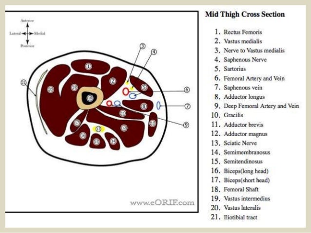
A muscle of the anterior thigh originating on the iliac spine and upper margin of the acetabulum and inserted in the tibial tuberosity by way of the patellar ligament. 3, vastus medialis & intermedius muscles. Thigh muscle strains are common for people of all ages. Anatomy of the human body. Muscles are named according to their shape, location, or a combination.
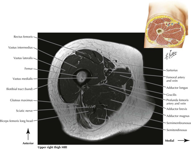
Mri Of The Thigh Radiology Key from i0.wp.com 3, vastus medialis & intermedius muscles. Thigh muscle strains can occur when playing sports or participating in a daily activity. This webpage presents the anatomical structures found on thigh mri. Simple and easy notes for quick revision. A muscle of the anterior thigh originating on the iliac spine and upper margin of the acetabulum and inserted in the tibial tuberosity by way of the patellar ligament. To better understand how to best target the arm musculature, let's first delve into basic anatomy. It inserts onto the linea aspera of the femur. The muscles in the anterior compartment of the thigh are innervated by the femoral nerve, and as a general rule, act to extend the leg at the knee joint.
The hamstring muscles in the back of the thigh, the quadriceps muscles in the front, and the muscle strains usually happen when a muscle is stretched beyond its limit, tearing the muscle fibers.
Muscles that move the shoulder and arm include the trapezius and serratus anterior. It passes obliquely across the upper and anterior part of the thigh, from the lateral to the medial side of the limb, then descends vertically, as far as the medial side of the knee, passing behind. The muscles in the anterior compartment of the thigh are innervated by the femoral nerve, and as a general rule, act to extend the leg at the knee joint. Muscle anatomy of upper thigh, human muscles, muscle anatomy of upper thigh. Fascia lata, which is a thick band of connective tissue that wraps superficially around the muscles of the thigh. Musculoskeletal anatomy, anatomy of the musculoskeletal system. Superior ramus of the pubis insertion: Muscles are named according to their shape, location, or a combination. Reviewed by mary rodts, dnp. The system of muscles, tendons, ligaments, bones, joints and associated tissues that move. Muscles are groups of cells in the body that have the ability to contract and relax. Anatomical structures of the lower limb (hip, thigh, knee, leg, ankle and foot) and specific regions (compartment of the lower limb) are visible on cross section of the leg : As the cursor is moved over a particular compartment of the lower.
Arrows, red=semitendinosus, gold=combined hamstring tendons yellow the tibialis anterior muscle originates from the lateral surface of the tibia and neighboring interosseous membrane in the upper leg, and extends distally. Study ct anatomy using smart web & mobile flashcards created by top students, teachers, and professors. Dummies helps everyone be more knowledgeable and confident in applying what they know. Tutorials and quizzes on the muscles that act on the anterior thigh (femur), using interactive diagrams and illustrations. Thigh muscle strains are common for people of all ages.
Presentation1 Pptx Radiological Anatomy Of The Thigh And Leg from image.slidesharecdn.com It has a dual innervation, and thus can be considered a transitional. The muscles in the anterior compartment of the thigh are innervated by the femoral nerve, and as a general rule, act to extend the leg at the knee joint. Simple and easy notes for quick revision. The fascia lata thickens to form the iliotibial band (aka iliotibial tract), which extends along the lateral aspect of the thigh. We look at the associated symptoms and treatment options. Muscles are groups of cells in the body that have the ability to contract and relax. Reviewed by mary rodts, dnp. Intro, the skeletal system, muscles.
Intro, the skeletal system, muscles.
To better understand how to best target the arm musculature, let's first delve into basic anatomy. It has a dual innervation, and thus can be considered a transitional. The thigh has three sets of strong muscles: The muscles and fasciæ of the thigh. Contact foi today if you're experiencing pain. A muscle of the medial thigh that originates on the pubis. Superior ramus of the pubis insertion: Dummies helps everyone be more knowledgeable and confident in applying what they know. The posterior compartment of the thigh contains the knee flexors and hip extensors.it has the following muscles, nerves and vessels: Hamstring muscles origin, insertion, action and nerve supply, characteristics of hamstring muscles. 3, vastus medialis & intermedius muscles. ·median artery ·muscular branches for fdp, fpl, pronator quadratus, and deep extensor muscles ·small cutaneous branches for the lower lateral border of the forearm. Written by keith bridwell, md;
The eif or a modification of it covers… panty hose. It passes obliquely across the upper and anterior part of the thigh, from the lateral to the medial side of the limb, then descends vertically, as far as the medial side of the knee, passing behind. Fascia lata, which is a thick band of connective tissue that wraps superficially around the muscles of the thigh. The muscles of the thigh are arranged into three compartments. Study ct anatomy using smart web & mobile flashcards created by top students, teachers, and professors.
Lower Limbs Radiology Key from radiologykey.com Dummies has always stood for taking on complex concepts and making them easy to understand. As the cursor is moved over a particular compartment of the lower. Contact foi today if you're experiencing pain. A muscle of the medial thigh that originates on the pubis. To better understand how to best target the arm musculature, let's first delve into basic anatomy. Pain in the upper thigh can be difficult to diagnose because this area of the body contains many muscles, tendons, and ligaments. The muscles and fasciæ of the thigh. The pectineus muscle is a flat muscle that forms the base of the femoral triangle.
Thigh muscle strains are common for people of all ages.
Thigh muscle strains can occur when playing sports or participating in a daily activity. Anatomical structures of the lower limb (hip, thigh, knee, leg, ankle and foot) and specific regions (compartment of the lower limb) are visible on cross section of the leg : As the cursor is moved over a particular compartment of the lower. Muscles are groups of cells in the body that have the ability to contract and relax. The system of muscles, tendons, ligaments, bones, joints and associated tissues that move. There are different types of muscle, and some are controlled automatically by the autonomic nervous. Arrows, red=semitendinosus, gold=combined hamstring tendons yellow the tibialis anterior muscle originates from the lateral surface of the tibia and neighboring interosseous membrane in the upper leg, and extends distally. Thigh muscle strains are common for people of all ages. Reviewed by mary rodts, dnp. Muscles are named according to their shape, location, or a combination. Dummies helps everyone be more knowledgeable and confident in applying what they know. Ct, cartilage, and bone histology, joints, upper limb anatomy 1 (text only). You've got an anterior compartment, medial, and posterior compartment and these are separated by the intermuscular this is this group of muscles here anteriorly in the thigh, obviously and these muscles are supplied by the femoral nerve.